BACKGROUND: The involvement of lipid metabolism in tumourigenesis and the progression of clear cell renal cell carcinoma (ccRCC) have been reported. However, the role of phospholipid profile alterations in ccRCC has not yet been systematically explored. In the present study, we compared the phospholipid compositions between ccRCC and paired normal renal tissues. METHODS: The phospholipid compositions of paired ccRCC and normal renal tissues were evaluated using liquid chromatography tandem mass spectrometry (LC/MS/MS). To evaluate the mRNA and protein levels of lysophosphatidylcholine acyltransferase (LPCAT), which converts lysophosphatidylcholine (LPC) to phosphatidylcholine (PC), qRT-PCR, western blotting and immunohistochemistry were performed. The correlations of LPCAT1 expression with clinicopathological features and prognosis were assessed. In addition, siRNAs were used to knockdown LPCAT1 expression in ccRCC cell lines, and its effect on cell proliferation, cell cycle, migration and invasion were investigated. RESULTS: The phospholipid compositions of ccRCC and normal renal tissues were significantly different. Multiple LPC species were decreased and corresponding PC species were increased in cancer tissues. The mRNA and protein levels of LPCAT1 were up-regulated in ccRCC tissues compared with normal renal tissues, and LPCAT1 expression was significantly correlated with unfavourable pathological features (higher tumour grade, higher TNM stage and larger tumour size) and overall survival. In cell line experiments, LPCAT1 knockdown depleted PCs, inhibited cell proliferation, migration and invasion and induced cell cycle arrest at the G0/G1 phase. CONCLUSION: Selective changes in PC and LPC composition were observed in ccRCC tissues. The overexpression of LPCAT1 promotes the development and progression of ccRCC, likely through the conversion of LPC to PC.
文章引用产品
-
-
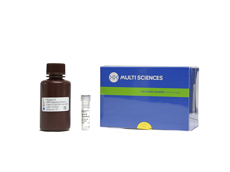
- CCS012
- 周期试剂盒
Cell Cycle Staining Kit 细胞周期检测试剂盒
-
¥390.00
-
-
- CCS012
- 周期试剂盒
Cell Cycle Staining Kit 细胞周期检测试剂盒
- ¥390.00
-
 Powered by Bioz