BACKGROUND: Stem cell-based therapy to treat liver diseases is a focus of current research worldwide. So far, most such studies depend on rodent hepatic failure models. The purpose of this study was to isolate mesenchymal stem cells from human placenta (hPMSCs) and determine their therapeutic potential for treating Chinese experimental miniature pigs with acute liver failure (ALF). METHODS: hPMSCs were isolated and analyzed for their purity and differentiation potential before being employed as the donor cells for transplantation. ALF models of Chinese experimental miniature pigs were established and divided into four groups: no cell transplantation; hPMSCs transplantation via the jugular vein; X-ray-treated hPMSCs transplantation via the portal vein; and hPMSCs transplantation via the portal vein. The restoration of biological functions of the livers receiving transplantation was assessed via a variety of approaches such as mortality rate determination, serum biochemical analysis, and histological, immunohistochemical, and genetic analysis. RESULTS: hPMSCs expressed high levels of CD29, CD73, CD13, and CD90, had adipogenic, osteogenic, and hepatic differentiation potential. They improved liver functions in vivo after transplantation into the D-galactosamine-injured pig livers as evidenced by the fact that ALT, AST, ALP, CHE, TBIL, and TBA concentrations returned to normal levels in recipient ALF pigs. Meanwhile, histological data revealed that transplantation of hPMSCs via the portal vein reduced liver inflammation, decreased hepatic denaturation and necrosis, and promoted liver regeneration. These ameliorations were not found in the other three groups. The result of 7-day survival rates suggested that hPMSCs transplantation via the portal vein was able to significantly prolong the survival of ALF pigs compared with the other three groups. Histochemistry and RT-PCR results confirmed the presence of transplanted human cells in recipient pig livers (Groups III, IV). CONCLUSIONS: Our data revealed that hPMSCs could not only differentiate into hepatocyte-like cells in vitro and in vivo, but could also prolong the survival time of ALF pigs. Regarding the transplantation pathways, the left branch of the portal vein inside the liver was superior to the jugular vein pathway. Thus, hPMSCs transplantation through the portal vein by B-ultrasonography may represent a superior approach for treating liver diseases.
文章引用产品
-
-
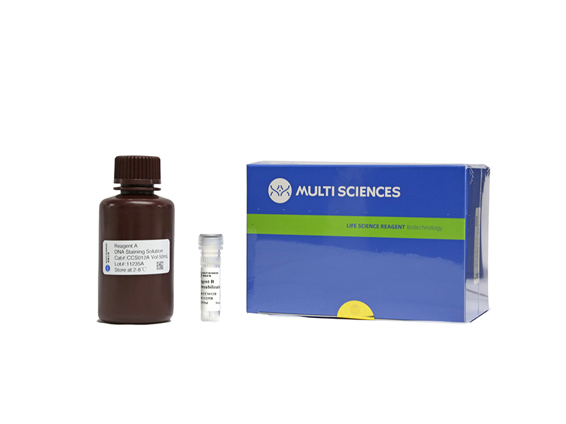
- CCS012
- 周期试剂盒
Cell Cycle Staining Kit 细胞周期检测试剂盒
-
¥390.00
-
-
- CCS012
- 周期试剂盒
Cell Cycle Staining Kit 细胞周期检测试剂盒
- ¥390.00
-
 Powered by Bioz