https://www.liankebio.com/article-information_Tech-2516.html
作者:联科生物 发布日期:2018-12-12 09:00
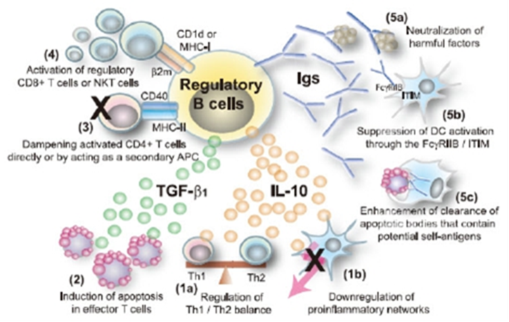
| ? 背景知识简介 对病原体产生免疫并将其清除,通常需要 B 细胞依赖的体液免疫应答和T细胞依赖的细胞免疫应答。B 细胞可通过分泌抗原特异性的抗体以及辅助诱导CD4+T 细胞活化来正向调控免疫应答。然而近来发现 B 细胞中有一群特异性的亚群可负向调控免疫应答,称为调节性 B 细胞,即 Breg。Breg 的缺失会加剧病症,包括炎症、自身免疫疾病(如接触性超敏反应(CHS)、实验性自身免疫性脑脊髓炎(EVE)、慢性肠炎和胶原酶诱导的关节炎(CIA))、癌症和感染疾病等[1-6]。 Breg 的免疫应答调节机制包括:1. 分泌 IL-10 调节 Th1/Th2 平衡(1a)、直接抑制炎症级联反应(1b);2. 分泌 TGF-β1 诱导效应 T 细胞凋亡;3. 直接或作为二级 APC 抑制活化的 CD4+T 细胞;4. 以 β2 微球蛋白依赖的方式(MHC I 和 CD1d)招募 Treg 亚群(CD8+T 细胞和 NKT 细胞);5. 分泌 IgG 和 IgA 中和有害可溶性因子(5a)、通过 IgG/FcγRIIB 抑制 DC/巨噬细胞的活化(5b)以及增强清除含有潜在自身抗原的凋亡细胞(5c)(图 1)。
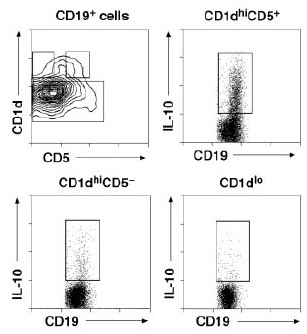
最近发现一群调节性 B 细胞的亚群,通过 IL-10 来调节 T 细胞依赖的 CHS 和EVE。该 B 细胞亚群的表型是独一无二的 CD1dhiCD5+CD19hi[5],在野生型小鼠的脾脏中约占 1-2%。在体外经 LPS、PMA、ionomycin 和 monensin 刺激后,可诱导产生分泌免疫抑制性细胞因子 IL-10 的 CD1d hiCD5+ B 细胞,称为 B10 细胞,以区分于其它调节性 B 细胞亚群。去除 B10 细胞可能增强某些免疫反应;增强 B10细胞功能可抑制一些自身免疫性疾病(如接触性过敏反应)中的炎症和过程的免疫反应。 IL-10 对大多数造血细胞具有抗炎症和抑制作用。IL-10 不仅可抑制 Th1 和 Th2细胞的极化[8, 9],也能抑制单核细胞和巨噬细胞分泌促炎性因子[10]。IL-10-/-小鼠会产生炎性肠炎,也会加剧 EVE、CIA 和 CHS[11]。因此 IL-10 可抑制炎症和自身免疫应答。B10 细胞不是 IL-10 调控的唯一来源,因此在炎症过程中需要进一步阐明 B10 和调节性 T 细胞的各自功能。 但 IL-10 在肿瘤免疫中的作用仍有争议。IL-10 可促进肿瘤生长,抑制免疫效应细胞的功能。然而 IL-10 可促进 CD8+T 细胞和 NK 细胞对肿瘤排斥,而且还能抑制肿瘤诱导的血管发生,并增强肿瘤毒性分子如 NO 的分泌,导致肿瘤退化。 有学者认为调节性 B 细胞也许可以分为“先天型”(多克隆刺激诱导 IL-10 分泌)和“获得型”(抗原特异性 IL-10 分泌)。LPS 的刺激可使 B10 分泌 IL-10;而调节性 B 细胞分泌 IL-10 也需要 CD40 的参与。 Breg 在炎症条件下被特异性地诱导,能够抑制炎症反应的加剧和/或加速恢复进程。但目前仍然有许多有关调节性 B 细胞的问题需要进一步的研究阐明。如Breg 是否具有抗原特异性?支持调节性 B 细胞和致病性 B 细胞发育的条件是什么?调节性 B 细胞是否也具有类似调节性 T 细胞的特异性转录标志如 FoxP3?调节性 B 细胞在某些炎症条件下是否具有致病性?或者其仅具有调节性功能?调节性功能(分泌 IL-10、TGF-β 和二级 APC 活性)是由同一群 Breg 还是不同 Breg 亚群发挥的?Breg 活化的信号通路是什么?Breg 的存在说明具有不同的机制来调控免疫应答,其中 B10 在自身免疫疾病和炎症反应中发挥了重要的调控作用。 PMA Multisciences,CS0001,溶于乙醇,0.1 mg/ml,100μl 包装。-20℃保存。 Ionomycin Multisciences,CS0002,溶于乙醇,1 mg/ml,50μl 包装。-20℃保存。 Monensin Multisciences,CS0004,溶于乙醇,50 mg/ml,100μl 包装。4℃保存至少4个月。 LPS 小鼠:Anti-Mouse CD16/CD32 (2.4G2), Purified (Multisciences, AM016) CD19 MicroBeads, human (99-130-050-301),检测抗体如 Anti-Human CD19 APC(eBioscience, 85-17-0199) 或 Anti-Human CD19 FITC (eBioscience, 85-11-0199) 或Anti-Human CD19 PE (eBioscience, 85-12-0199)等 小鼠: CD19 MicroBeads, mouse (Miltenyi, 99-130-052-201),检测抗体如 Anti-Mouse CD19 FITC (eBioscience, 85-11-0193) 或 Anti-Mouse CD19 PE (eBioscience, 85-12-0193)或Anti-Mouse CD19 APC (eBioscience, 85-17-0193)等;或 CD45R (B220) MicroBeads, mouse (Miltenyi, 99-130-049-501),检测抗体如Anti-Human/Mouse CD45R (B220) FITC (eBioscience,85-11-0452)或Anti-Human/Mouse CD45R (B220) PE (eBioscience, 85-12-0452)或Anti-Human/Mouse CD45R (B220) APC (eBioscience, 85-17-0452)等
小鼠:
八、流式染色缓冲液 工作液配制:将贮存液 1:100 稀释于 RPMI 1640 或 PBS (无菌,无 Na3N),此时为 1μg/ml 工作浓度:50 ng/ml 细胞培养:每 100μl 培养体系中加入 5μl 工作液 2. Ionomycin (Multisciences, CS0002) 工作液配制:贮存液 1:20 稀释于 RPMI 1640 或 PBS (无菌,无 Na3N),此时为50μg/ml 工作浓度:500 ng/ml 细胞培养:每 100μl 培养体系中加入 1μl 工作液 3. Monensin (Multisciences, CS0004) 工作浓度:2μM 细胞培养:每 100μl 培养体系中加入 2.78μl 贮存液 4. LPS 工作浓度:10μg/ml a. 外周血 1. 采集至少 2ml 抗凝血,4 小时内使用 2. 加入等量的缓冲液(如 PBS, pH 7.2-7.4)稀释血液 3. 取与稀释后血液等体积的淋巴细胞分离液加入至离心管 4. 将稀释血液缓慢加入至淋巴细胞分离液上,保持两者的界面清晰 5. 室温 300g 离心 20min 6. 吸取单个核细胞层,转移至新的离心管中,加入缓冲液,300 g 离心 10 min,弃上清后,再洗涤一次 7. 弃上清,用细胞培养液重悬,台盼蓝染色计数活细胞(活细胞比例应大于 95%) 8. 调整细胞浓度为 2×10⁶ 个/ml b. 脾脏 1. 将脾脏切成小块后,经 400 目铜网研磨,制成单细胞悬液 2. 取与脾细胞悬液等体积的淋巴细胞分离液加入至离心管 4. 将脾细胞悬液缓慢加入至淋巴细胞分离液上,保持两者的界面清晰 5. 室温 300 g 离心 20min 6. 吸取单个核细胞层,转移至新的离心管中,加入缓冲液,300 g 离心 10 min,弃上清后,再洗涤一次 7. 弃上清,用细胞培养液重悬,台盼蓝染色计数活细胞(活细胞比例应大于 95%) 8. 调整细胞浓度为 2×10⁶ 个/ml B. 磁珠分选纯化法(推荐,可选) 以 CD19 MicroBeads, human (130-050-301)分选纯化人 B 细胞[7]。或以 CD19 MicroBeads, mouse (Miltenyi, 130-052-201)或 CD45R (B220) MicroBeads, mouse (Miltenyi, 130-049-501)进行分离纯化小鼠 B 细胞[5, 12, 13]。并以抗人 CD19 抗体或抗小鼠 CD19 抗体或抗小鼠 CD45R 抗体进行流式检测。具体操作请参照说明书。三、细胞培养: 淋巴细胞分离液分离的单个核细胞或磁珠分选纯化的 B 细胞(1×10⁶ 个/ml)接种 于 细 胞 培 养 板 中 , 刺 激 组 加 入 LPS(10μg/ml) 、 PMA(50ng/ml)、 ionomycin (500ng/ml)和 monensin (2μM),37℃,5%CO2 恒温细胞培养箱中孵育 5h。未刺激组不加 LPS,其余同。对于同时检测 B10 和 B10 细胞祖细胞(B10Pro cell)的实验,需培养 48h,使 B10Pro 细胞发育为成熟的 B 细胞,在最后 5h 再加入上述刺激剂和阻断剂[14]。四、流式检测(四色法) 1. 细胞计数,取未刺激(A 管)和刺激(B 管)的细胞(2×10⁵-2×10⁸/管),加入适量(具体参照说明书)FcR 阻断剂(Human Fc Receptor Binding Inhibitor Purified(人)或 Anti-Mouse CD16/CD32 Purified(小鼠)), 加入 Staining Buffer 补充至 100μl,冰上孵育 20min。 2. 将 A 管和 B 管分别等分成 2 份,标为 A1(同型对照管)、A2(样品管)、B1(同型对照管)、B2(样品管)。 3. 各管按下表加入相应的抗体(具体量请参照说明书): A. 人
B.小鼠
每管加入 Staining Buffer 补充至 100μl,室温避光孵育 15min。 4. 每管加入 1-2ml Staining Buffer,300g 离心 5min,弃上清。 5. 每管加入 100μl Fix&Perm 中的 Reagent A(即固定液),室温避光孵育 15min。 6. 每管加入 3ml Staining Buffer,300g 离心 5min,弃上清。 7. 每管加入 100μl Fix&Perm 中的 Reagent B(即破膜和溶血液),同时各管按下表加入相应的抗体(具体量请参照说明书): A. 人
B.小鼠
室温避光孵育 15min。 8. 每管加入 3ml Staining Buffer,300g 离心 5min,弃上清。 9. 每管加入 0.5ml Staining Buffer,重悬细胞,上机检测
【参考文献】 2. Fillatreau, S., et al., B cells regulate autoimmunity by provision of IL-10. Nat Immunol, 2002. 3(10): p. 944-50. 3. Mizoguchi, A., et al., Chronic intestinal inflammatory condition generates IL-10-producing regulatory B cell subset characterized by CD1d upregulation. Immunity, 2002. 16(2): p. 219-30. 4. Mauri, C., et al., Prevention of arthritis by interleukin 10-producing B cells. J Exp Med, 2003. 197(4): p. 489-501. 5. Yanaba, K., et al., A regulatory B cell subset with a unique CD1dhiCD5+ phenotype controls T cell-dependent inflammatory responses. Immunity, 2008. 28(5): p. 639-50. 6. Matsushita, T., et al., Regulatory B cells inhibit EAE initiation in mice while other B cells promote disease progression. J Clin Invest, 2008. 118(10): p. 3420-30. 7. Mizoguchi, A. and A.K. Bhan, A case for regulatory B cells. J Immunol, 2006. 176(2): p. 705-10. 8. Asseman, C., et al., An essential role for interleukin 10 in the function of regulatory T cells that inhibit intestinal inflammation. J Exp Med, 1999. 190(7): p. 995-1004. 9. Grunig, G., et al., Interleukin-10 is a natural suppressor of cytokine production and inflammation in a murine model of allergic bronchopulmonary aspergillosis. J Exp Med, 1997. 185(6): p. 1089-99. 10. Riley, J.K., et al., Interleukin-10 receptor signaling through the JAK-STAT pathway. , 1999. 274(23): p. 16513-21. 11. Bettelli, E., et al., IL-10 is critical in the regulation of autoimmune encephalomyelitis as demonstrated by studies of IL-10- and IL-4-deficient and transgenic mice. J Immunol, 1998. 161(7): p. 3299-306. 12. Matsushita, T., et al., Regulatory B cells (B10 cells) and regulatory T cells have independent roles in controlling experimental autoimmune encephalomyelitis initiation and late-phase immunopathogenesis. J Immunol, 2010. 185(4): p. 2240-52. 13. Yanaba, K., et al., The development and function of regulatory B cells expressing IL-10 (B10 cells) requires antigen receptor diversity and TLR signals. J Immunol, 2009. 182(12): p. 7459-72. 14. Iwata, Y., et al., Characterization of a rare IL-10-competent B-cell subset in humans that parallels mouse regulatory B10 cells. Blood, 2011. 117(2): p. 530-41. 15. Bouaziz, J.D., K. Yanaba, and T.F. Tedder, Regulatory B cells as inhibitors of immune responses and inflammation. Immunol Rev, 2008. 224: p. 201-14. |